

![]()
![]()
![]()
![]()
![]()
![]()
![]()
![]()

![]()
|
Research in the Simpson Group |
|
|
Chemical Imaging over Wavelength and Time |
|
|
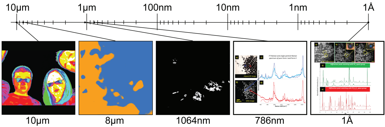
Spatial arrangements often define function, but are challenging to access by conventional sample-averaging measurement approaches. Localizing chemical characterization to locations only a few ?m in diameter can greatly increase the signal to noise and recover information on the spatial correlations between composition in samples ranging from tissue sections to pharmaceutical formulations. Nonlinear optical methods using ultrafast laser sources can provide selective image contrast based on symmetry, complemented by spectroscopic/diffraction analyses at highly localized regions of interest. Pairing linear and nonlinear optical imaging modalities enables high confidence in chemical classification with high speed and high spatial resolution. |
|
|
|
|
Integrated Imaging |
|
|
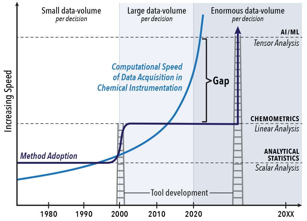
The Moore's Law increase in information content accessible from ever-increasing processing speeds enables the rapid acquisition of enormous volumes of chemical imaging data. Coupling this tsunami of information with data mining and machine learning algorithms into the data acquisition pipeline opens up opportunities lost in more conventional acquire-first, analyze-later strategies. This integrated imaging in which advanced algorithms are embedded into the design and implementation of the hardware have greatly enhanced the frame rates achievable in beam-scanning imaging, have enabled merging of information from multiple disparate platforms, and greatly increased the number of accessible wavelength channels in hyperspectral image acquisition. |
|
|
|
|
Composition |
|
|
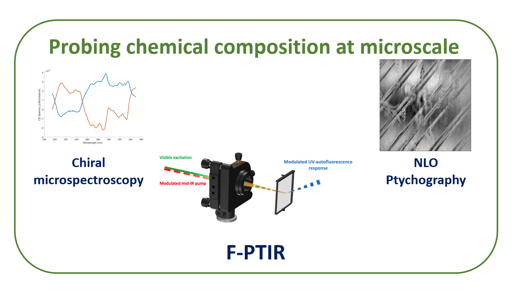
Exciting recent developments in optical microscopy techniques enabled mapping the morphology of samples with unprecedented lateral resolution approaching atomic scales. However, many of these techniques are unable to recover the chemical composition of the studied systems, which might be critical in studying heterogeneous systems such as cells of pharmaceutical formulations. In Simpson's lab, we are constantly working on developing new approaches to chemically-specific analysis using such strategies as chiral-specific spectroscopy, phase imaging, and photothermal microscopy. We have recently demonstrated a new approach to performing sub-diffraction mid-IR microspectroscopy that relies on detecting subtle temperature-induced changes in fluorescence quantum intensity caused by local absorption of mid-IR radiation. Link to the C&E News feature article on Fluorescence-detected Photothermal mid-IR microscopy (F-PTIR) |
|
|
|
|
Dynamics |
|
|
Quantifying mobility of small-molecules and proteins in complex heterogeneous matrices is critical to predicting stability and potential bioavailability. Fluorescence recovery after photobleaching is a simple technique to measure diffusivity at a single spot by photobleaching and subsequent fluorescence recovery. To interrogate more complex samples, patterned photobleaching followed by spatial Fourier transform allows quantification of diffusion at a series of discrete length scales simultaneously, while sampling the full field of view. In addition, analysis at multiple length scales allows interrogation of recovery contributions from bulk diffusion vs. interfacial exchange/diffusion. |
|
|
|
|
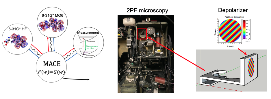
Computation |
|
|
MACE (multi-agents consensus equilibrium) aims to fuse multiple descriptions for one consensus result at equilibrium to bridge computations with measurements. |
|
|
|
|
Previous Research |
|
|
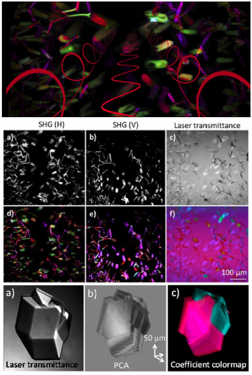
Second Harmonic Generation Microscopy |
|
|
Second order nonlinear optical effects, including second harmonic generation (SHG) and sum-frequency generation (SFG) enable image contrast directly tied to the local symmetry and molecular ordering in biological assemblies. We take advantage of both the overall intensity for image contrast and the exquisite sensitivity to polarization-dependence to characterize both structure and structural evolution in biological systems. Of particular interest are efforts to detect protein crystal formation to support macromolecular structure determination, small-molecule crystal formation of active pharmaceutical ingredients to support stability assessments, and tissue imaging to provide fundamental insights into native biopolymer organization. Additionally, we are using SHG microscopy to target subsequent measurement by Raman and synchrotron X-ray diffraction, combining the fast imaging capabilities of SHG with chemical information-rich orthogonal techniques. |
|
|
|
|
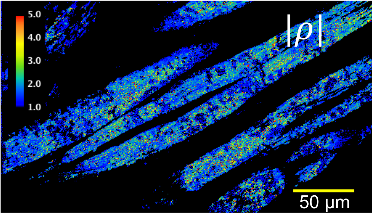
Polarization-Dependent Imaging |
|
|
The information content within an SHG measurement can be enhanced through polarization-dependent imaging. By modulating the incident polarization and measuring the intensity of the polarized output, structural characteristics of a sample can be recovered. Our current focus is using fast (8MHz) polarization modulation to enable video-rate polarization-dependent imaging. Such methodology could be used to rapidly characterize structure within many systems, potentially allowing such applications as disease monitoring in collagenous fibers, identification of twinning in crystals, or real-time discrimination of crystal polymorphic form. |
|
|
|
|
Colormap of Polarization-dependent Crystal Imaging |
|
|
A brief introduction of the instrument and algorithm development for nonlinear optical Stokes ellipsometric microscopy and its application in the analysis of pharmaceutical ingredients and biological tissues. Made by Dr. Ximeng You Dow for 2015 American Chemical Society meeting at Boston. |
|
|
Triboluminescence |
|
|
The pharmaceutical industry has become a victim of it's own rapid success. The improvement of drugs with respect to target specificity comes with a corresponding decrease in water solubility because of the increase of molecular complexity. The pharmaceutical industry is increasingly formulating it's active ingredients as amorphous solid dispersions (ASDs) to increase the apparent solubility. ASDs have their own problems, with the active ingredient being in it's amorphous form, it is thermodynamically favorable to recrystallize. We are using Triboluminescence as a way to measure the crystallinity within amorphous solid dispersions. We have built a custom instrument to measure crystallinity with the help of the Jonathan Amy Facility for Chemical Instrumentation. Using this relatively inexpensive instrument we have demonstrated a limit of detection of 140 ppm, which is a large improvement over the 1-2% limit of detection for commonly used instruments (PXRD, DSC, ssNMR, etc.) For more information please read our recently published article at Analytical Chemistry here. |
|
|
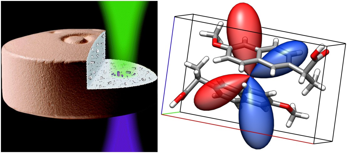
Mueller Tensors for Nonlinear Optics in Turbid Media |
|
|
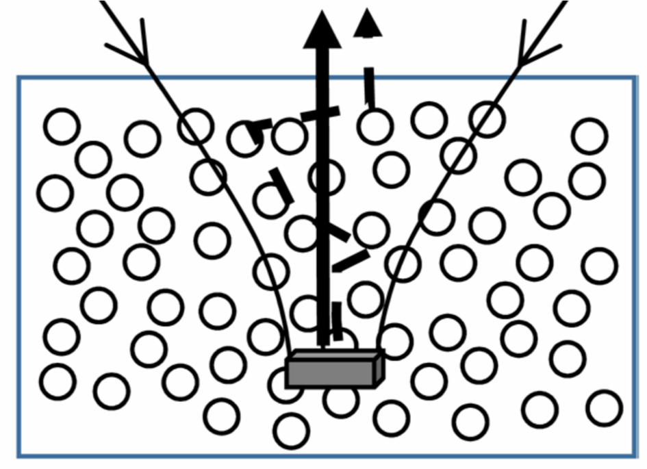
Turbidity and scattering can alter polarization state, introducing uncertainty in the measurement and interpretation of polarization-dependent optics. The Jones formalism for polarization-state analysis is incapable of representing the electric field with no defined polarization state and is thus insufficient as a formalism for interpretation of polarization-dependent measurements with significant contributions of intensity from unpolarized light. The Stokes-Mueller formalism is a more general approach to defining polarization of light via the field intensity, however it comes at a cost of greater complexity as the base vector for a single photon contains 4 elements, twice as large as the Jones vectors' 2. Recently, we developed a framework which connects the Jones framework directly to the Stokes framework, allowing predictive capacity for SHG behavior in the highly depolarized regime, such as when imaging thick tissue sections. This work leverages the community-wide usage of the simplest representation of the nonlinear optical susceptibility, the Jones tensor, while maintaining the generality of the Stokes framework. We have demonstrated the validity of the framework for interpretation of SHG polarization dependence from an unpolarized source, as measured by introducing a depolarizing optic into the Fourier plane of a beam-scanning microscope. Our theory agrees well for experimental determination of the polarization dependence of SHG from Z-cut quartz with a depolarized source. Furthermore, the depolarizing optic in the Fourier plane was also used as a method to recover orientation and tensor element magnitudes for thin collagenous tissue sections. Most recently, we have demonstrated the imaging of the unique tensor elements of collagen within a 40 ?m thick section of mouse tail tendon. |
|
|
Contact Webmaster: Yechan Hwang. Visit our Website Development Board for more information.
Copyright © 2016, Simpson Research Group, Department of Chemistry, Purdue University, all rights reserved
560 Oval Drive, West Lafayette, IN 47907