Purdue chemists use mass spec tool to help surgeons detect tumor tissue during cancer surgery
2024-05-28
Writer(s): Steve Scherer
Researchers from Purdue and Mayo Clinic were able to quickly and accurately identify glioma mutations in tumor tissue during brain cancer surgery, according to a recent study published in the Proceedings of the National Academy of Sciences (PNAS).
During surgeries at Mayo Clinic Florida, doctors resected brain tumor tissue and provided small tumor sample smears to Purdue chemists who quickly analyzed them using a mass spectrometer.
“We are measuring 2-hydroxyglutarate or 2HG which is a specific biomarker that is only present in IDH-mutant glioma. Glioma is a prevalent form of brain tumor, and some gliomas carry a mutation. For the surgeons, knowing whether the tumor carries the mutation is important for diagnosis, prognosis, and treatment of glioma patients,” explained Mahdiyeh Shahi, a Purdue chemistry graduate student who helped analyze the tissue samples during several of the surgeries at the Jacksonville, Fla. hospital.
“The standard of care at the moment is that during surgery, there is no information obtained with regard to molecular characterization of the tumors – whether mutant or not mutant,” said R. Graham Cooks, Henry Bohn Hass Distinguished Professor of Chemistry.
Shahi says postoperative molecular analysis of tissue is the current standard of care, and even that can take hours or days in some instances.
“Our measurements and prediction can be done in less than two minutes, which makes it ideal for an interoperative diagnostic tool to be used during the surgery which could give more information to the surgeon about clear margins, so they can make better decisions on removing tumor tissue and when to stop removing it,” she explained.
“This gives the surgeons a chemical label as a guide to where there is tumor tissue. The mutation labels the tissue – with mutant metabolite molecules – specifically with a small molecule called 2HG and we find it very easily,” added Cooks, whose research lab has been involved in brain tumor analysis for more than a decade.
An independent team of Chinese researchers from Tsinghua University and Huashan Hospital coauthored the PNAS paper, where that team performed similar measurements using a different mass spectrometer.
According to the study, none of the 31 patients at Mayo Clinic or the 74 patients at Huashan Hospital were misclassified when analyzing the tumor core biopsies.
“It’s amazing to get one hundred percent accuracy. We know where the tumor is every time we get 2HG. One disadvantage is it is not applicable to all gliomas - most tumors are not mutant. But the advantage is that is amazingly specific,” said Cooks.
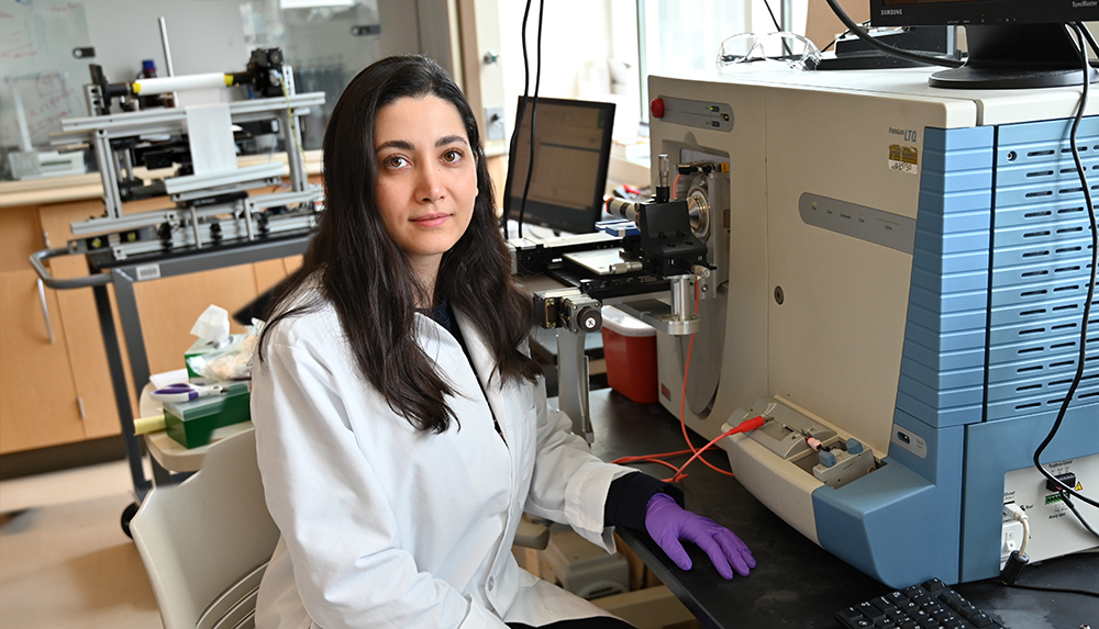
Mahdiyeh Shahi, a third-year chemistry graduate student from Tehran, Iran, using a mass spectrometer in R. Graham Cooks' analytical chemistry lab inside the Hall for Discovery and Learning Research located in Purdue University's Discovery Park. (Photo by Steve Scherer)