Mass spectrometry offers faster look at the edges of brain tumors
2026-10-01
Brain cancer margins can be especially difficult to map. Using mass spectrometry, researchers from Purdue University and the Mayo Clinic can map margins in resected tissue in a three-minute measurement as seen in the illustration above. Original illustration created by Mahdiyeh Shahi/Purdue University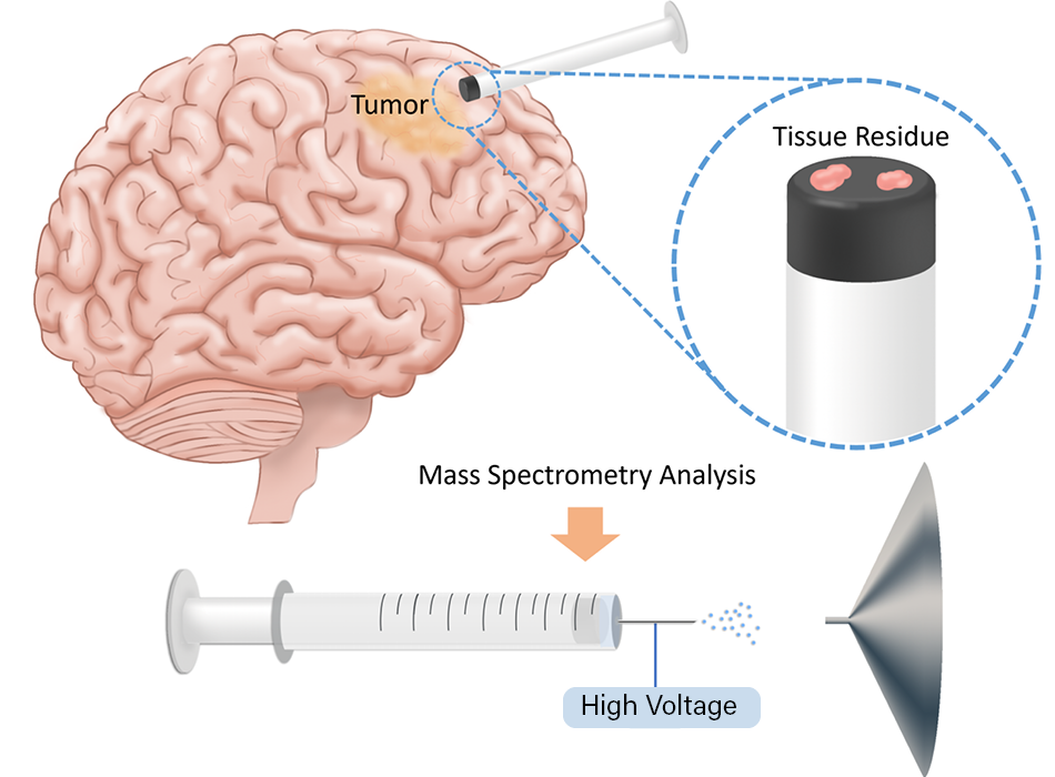
Glioma brain tumors are diffuse, so surgeons usually cannot excise all of the cancer during surgery. Currently, surgeons remove as much of the tumor as they can and then analyze the borders of the tumor after surgery. Researchers at Purdue University have developed a new method in which mass spectrometry could be used during surgery to evaluate tissue. This information could help guide the surgical team’s decisions, increasing the amount of cancerous tissue they are able to remove and improving the patient’s outcomes.
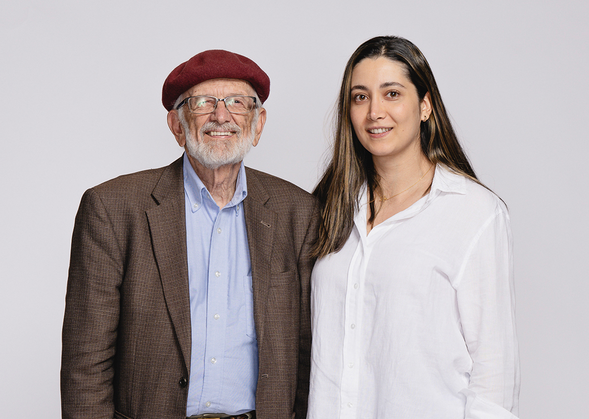
This research, which was recently published in PNAS, was led by Mahdiyeh Shahi, a former graduate student at Purdue University, and R. Graham Cooks, the Henry Bohn Hass Distinguished Professor of Chemistry in the James Tarpo Jr. and Margaret Tarpo Department of Chemistry at Purdue. Shahi is now a clinical chemistry postdoctoral fellow at the University of Minnesota Medical School. Shahi and Cooks worked in collaboration with noted brain surgeon Dr. Alfredo Quinones-Hinojosa of the Mayo Clinic; Quinones-Hinojosa’s postdoctoral fellow, Loizos Michaelides; and others from Mayo to develop and test this mass spectrometry method.
Gliomas are highly infiltrative and deadly brain tumors with complex molecular characteristics that complicate diagnosis and treatment. Brain surgery is one of the most important treatments for glioma and can strongly affect a patient’s prognosis and survival. Because glioma cells can spread into nearby healthy brain tissue, particularly at the tumor margins, they are difficult to detect using conventional technologies available in the operating room. Tumor tissue that remains in the brain after surgery can contribute to recurrence and make further treatment more challenging.
“The information we are getting is currently not available under standard of care procedures,” said Shahi. “If our diagnostic method were to be approved, it would provide actionable molecular information during surgery. This information is currently not available on the time scale (some hours) of the surgery. This information could directly influence treatment, including the extent of tumor resection, although the final surgical decision would be made by the surgeon based on the overall clinical context rather than the mass spectrometry results alone. It could also help guide the selection of targeted therapies.”
During brain tumor surgery, a small number of samples are taken from a surgeon-defined position in the brain for pathology. In this study, fractions of such samples were transferred onto the plunger of a hypodermic needle. Scientists would then spray a solution that measures the ratio of signals for multiple pairs of endogenous compounds in the brain using tandem mass spectrometry in a process that takes roughly three minutes.
“The study used 86 human brain samples, and the researchers showed that ratios of endogenous metabolites in brain tissue can distinguish glioma from non-tumor tissue with high confidence,” said Cooks. “It can also classify tumor subtypes and mutation status. The simple ambient ionization mass spectrometry approach enables fast, clinically compatible analysis with potential to improve margin assessment and enhance patient outcomes.”
Mass spectrometry is an analytical laboratory technique that measures the mass-to-charge ratio of ionized molecules to identify and quantify complex chemical compounds.
“The mass spectrometry technology we used was invented at Purdue,” said Cooks. “The method was based on ambient ionization making ions from molecules in the open environment. This allows molecular measurements to be made in situ. The first such method was described in 2004 and has since been commercialized and widely used. The new method used in this study — syringe touch spray — uses an ordinary hypodermic to take a sample from the patient and perform mass spectrometry analysis.”
The samples analyzed in this study were taken using MRI-guided precision surgery. The tissue samples were characterized by standard methods, from which molecular information is available only after surgery.
Now that this has been tested on 86 human samples, the team will explore the next steps to get this technique into surgical situations and diagnoses. A similar 50-plus patient study will need to be undertaken in which the technique is used during surgery. According to Cooks, this would pave the way for a clinic trial to be proposed.
“In previous work, our measurements have been made in the hospital, during surgery (PNAS 2024),” said Cooks. “In this case, we recorded the mass spectrometry remotely and compared with the available clinical information. The very simple and quick measurement has high discriminating power, distinguishing diseased from non-diseased tissue as well as providing information on tumor grade and mutation status. This technology would not replace the current standard of care during brain tumor surgery. Instead, it could provide surgeons with molecular information that is not currently available in the operating room, helping to guide surgical decisions during tumor removal, particularly at the tumor margins.”
The research team included Shahi and Cooks, along with Quiñones-Hinojosa, a professor of neurosurgery at Mayo Clinic in Jacksonville, Florida; Michaelides; Diogo Moniz Garcia, a former postdoctoral fellow at Mayo Clinic and current PGY3 neurosurgery resident at Washington University in St. Louis; and Aleeshba N. Basil of the Mayo Clinic. Kenneth Lane Virgin of Indiana University School of Medicine also contributed and was previously affiliated with Purdue’s Aston Laboratories for Mass Spectrometry. Cooks is a member of the Purdue Institute for Cancer Research and an Analytical Chemistry faculty in Purdue University’s College of Science.
This research was supported by the Purdue University Institute for Cancer Research (the National Institutes of Health grant P30CA023168), the Innovative Molecular Analysis Technologies (IMAT) Program of the National Cancer Institute at the NIH (R33CA240181-01A1), the National Center for Advancing Translational Sciences (NCATS), the New Chemistries for Undrugged Targets through ASPIRE (A Specialized Platform for Innovative Research Exploration) Collaborative Research Program (UG3TR004139), as well as corporate support from Waters Corporation. The specimens utilized in this investigation were established from invaluable patient-derived specimens collected through the Mayo Clinic Florida Neurosurgery BRIDGE Biobank, a central nervous system bioresource that facilitates translational research and advances scientific discovery on a global scale.
About Purdue Chemistry
The James Tarpo Jr. and Margaret Tarpo Department of Chemistry is internationally acclaimed for its excellence in chemical education and innovation, boasting two Nobel laureates in organic chemistry, the No. 1-ranked analytical chemistry program and a highly successful drug discovery initiative that has generated hundreds of millions of dollars in royalties. Learn more about chem.purdue.edu.
About Purdue University
Purdue University is a research institution ranked among the top 10 public universities in the United States. More than 111,000 students study at Purdue across multiple campuses, including more than 57,000 at our main campus locations in West Lafayette and Indianapolis. As a land-grant university committed to affordability and accessibility, Purdue’s main campus has frozen tuition 14 years in a row, enabling more students than ever to graduate debt-free.
Written by: Cheryl Pierce, College of Science
Contributors:
Mahdiyeh Shahi, former graduate student of Purdue University and current clinical chemistry postdoc fellow at the University of Minnesota Medical School
Graham Cooks, the Henry Bohn Hass Distinguished Professor of Chemistry in the James Tarpo Jr. and Margaret Tarpo Department of Chemistry
Photos by: Charles Jischke
Illustration provided by: Mahdiyeh Shahi