Simpson Group: Protein Crystal Detection Research
2009-12-21

Associate Professor Analytical & Physical Chemistry
Professor Garth Simpson’s research group is developing new methods to detect the formation and structure of membrane protein crystals―research that has applications in guiding rational drug design.
The difficulties in obtaining high resolution structures of integral membrane proteins have frustrated rational drug design efforts for these critical proteins, which comprise more than half of new drug targets. High-resolution structures of large proteins are most commonly generated from X-ray diffraction of protein crystals, with the major bottleneck often lying in the generation and detection of diffraction quality crystals.
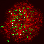
In this study, second harmonic generation (SHG) microscopy was used to detect membrane protein crystals in lipidic mesophase environments. By nature of the intrinsic chirality (i.e., molecular handedness) of proteins, allprotein crystals necessarily fall into crystal space groups that are bulk-allowed for SHG, or the frequency doubling of light. However, disordered assemblies of the same proteins, proteins in solution, and most crystals of simple salts typically produce no coherent SHG. As a result, SHG imaging is remarkably selective for protein crystals, enabling sensitive detection of protein microcrystals even in complex and highly scattering crystallization media such as the lipidic mesophases used in these studies.
This work involved collaboration between the Simpson research group, Prof. Vadim Cherezov at Scripps, the Chemistry Laser Laboratory and the Jonathon Amy Facility for Chemical Instrumentation.
Related links:
Analytical Chemistry article,
Simpson Research Group